Tuesday, October 31, 2006

Referred pain

Updated July 2009

If you woke up with a pain in your shoulder, you'd probably think something was wrong with your shoulder, right? Maybe you slept on it the wrong way, maybe you're a weekend warrior who threw the football a few too many times. In most cases, your hunch is probably right. Pain in the shoulder usually indicates an injury or disease that affects a structure in your shoulder, such as, say, your subacromial bursa or a rotator cuff tendon. Makes sense, doesn't it?

But you might be way off. Sometimes the brain gets confused, making you think that one part of the body hurts, when in fact another part of the body, far removed from the pain, is the real source of trouble. This curious (and clinically important) phenomenon is known as referred pain. For example, it's unlikely but possible that your shoulder pain is a sign of something insidious happening in your liver, gall bladder, stomach, spleen, lungs, or pericardial sac (the connective tissue bag containing the heart). Strange, isn't it? Conditions as diverse as liver abscesses, gallstones, gastric ulcers, splenic rupture, pneumonia, and pericarditis can all cause shoulder pain. How is that possible?

Neuroscientists still don't know precisely which anatomical connections are responsible for referred pain, but the prevailing explanation seems to work pretty well. In a nutshell, referred pain happens when nerve fibers from regions of high sensory input (such as the skin) and nerve fibers from regions of normally low sensory input (such as the internal organs) happen to converge on the same levels of the spinal cord. The best known example is pain experienced during a heart attack. Nerves from damaged heart tissue convey pain signals to spinal cord levels T1-T4 on the left side, which happen to be the same levels that receive sensation from the left side of the chest and part of the left arm. The brain isn't used to receiving such strong signals from the heart, so it interprets them as pain in the chest and left arm.

So what about that shoulder pain? All of organs listed above bump up against the diaphragm, the thin, dome-shaped muscle that moves up and down with every breath. The diaphragm is innervated by two phrenic nerves (left and right), which emerge from spinal cord levels C3, C4, and C5 (medical students remember these spinal cord levels using the mnemonic, "C3, 4, 5 keeps the diaphragm alive"). The phrenic nerves carry both motor and sensory impulses, so they make the diaphragm move and they convey sensation from the diaphragm to the central nervous system.

Most of the time there isn't any sensation to convey from the diaphragm, at least at the conscious level. But if a nearby organ gets sick, it may irritate the diaphragm, and the sensory fibers of one of the phrenic nerves are flooded with pain signals that travel to the spinal cord (at C3-C5). It turns out that C3 and C4 don't just keep the diaphragm alive; neurons at these two spinal cord levels also receive sensation from the shoulders (via the supraclavicular nerves). So when pain neurons at C3 and C4 sound the alarm, the brain assumes (quite reasonably) that the shoulder is to blame. Usually that's a good assumption, but sometimes it's wrong.


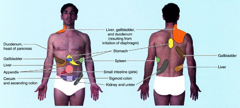
Illustration showing sites of referred pain from abdominal organs. From Moore and Dalley's Clinically Oriented Anatomy. Please note that I added the "tighty whities" with Photoshop (hey, this is a family friendly site).


Another example that seems bizarre until you know the anatomy is disease in the stomach causing pain between the shoulder blades. A classmate told me that one of her college professors complained to his doctor about pain in his upper back that wouldn't go away. It turns out that the professor had gastric cancer, a relatively aggressive and often incurable disease unless it's caught early. Unfortunately it wasn't caught early enough and it ended up taking his life. Maybe the outcome would have been different if the doctor had remembered that some of the nerve fibers to the stomach (specifically, visceral afferents that travel in the greater splanchnic nerve) convey pain signals to the same spinal cord levels (especially T5 and T6) that receive pain signals from the skin between the shoulder blades. This variety of referred pain is rare, but it happens often enough to be mentioned in anatomy textbooks.

Not all cases of referred pain are easy to find in textbooks. Take the appendix, for instance. That wormy little appendage of the colon is usually located in the right lower quadrant of the belly, nowhere near the diaphragm. Typically people with appendicitis feel diffuse pain or discomfort around the belly button, or sharp pain in the right lower quadrant if the appendix is getting ready to burst. But occasionally the pain refers to the right shoulder. Why? Note that I said the appendix is usually located in right lower quadrant. Sometimes, early in embryological development, the colon doesn't rotate as much as it should, and the appendix ends up in the right upper quadrant (or even on the left side of the belly). That malrotation isn't necessarily a problem for the patient until the appendix becomes swollen with infection, irritating both the diaphragm and the doctor who is trying to make a diagnosis!

So if your shoulder happens to hurt right now, should you be worrying about something bad in your belly? Probably not. The most common causes of shoulder pain are, by far, musculoskeletal problems like osteoarthritis, adhesive capsulitis, and rotator cuff tendinitis. Often, with the help of taking it easy and a few rounds of ibuprofen, the pain goes away on its own. But if you're still concerned, make an appointment with your family physician. Red flags include more ominous symptoms like fever, unexplained weight loss, and pain in the left shoulder or scapular area that gets worse with exertion.

Your doctor will want to know all about the pain. Was there a specific injury or did the pain come on slowly? Does the pain occur without movement of the shoulder? Can you make it worse with specific movements? Are there any other symptoms or chronic medical problems? The answers to these questions, along with a physical exam and appropriate imaging studies, will provide the information your doctor needs to understand the problem and create a treatment plan. The odds are good that the problem is relatively common and treatable. But if your doctor seems stumped, it wouldn't hurt to ask, "Is there any chance this could be referred pain?"

Sunday, September 17, 2006

Eye popping

I'm back!

Yes, Dear Reader, it's been too long, and I can't promise regular updates now that classes are in full swing, but I just couldn't pass up today's weird anatomy news flash:

Man sets sights on eye-popping record

Sun Sep 17, 7:08 AM ET

RIO DE JANEIRO, Brazil - Claudio Paulo Pinto is looking to break an eye-popping record. Literally. Pinto can pop his eyeballs out of their sockets at least 7 millimeters (0.3 inches), a national record for eye-popping according to RankBrasil, an organization modeled after the Guinness Book of World Records that lists Brazilian records.

A former driver, Pinto got a job scaring visitors in a commercial haunted house in Belo Horizonte, 210 miles north of Rio de Janeiro. But he recently was laid off, and now he seeks international recognition for his ability.

"I was measured by an opthamologist on television in January. I could pop my eyes out 7 millimeters," Pinto said by telephone Saturday. "Since then, my capacities have improved over 50 percent."

That could put Pinto close to the record. The title of "furthest eyeball popper" in the Guinness Book of World Records currently belongs to Kim Goodman of Chicago, who can pop her eyeballs 11 milimeters (0.43 inches) out of her sockets.

Pinto's ability is called "globe luxation." Doctors say it can strain blood vessels and nerves between the eyes and the head and feels unpleasant but usually doesn't cause lasting damage.

Pinto says he's been luxating his globes since he was 9 years old and "it doesn't hurt a bit."


How bizarre is that? At first I didn't think it was anatomically possible, but seeing is believing. I first learned about the phenomenon when I read this blog entry by Dr. Bill Lloyd, an ophthalmologist in Sacramento, California. Dr. Lloyd notes that several of his patients have been able to pop their eyes, and that there "have been instances where the eyelids have slipped behind the eyeball and trapped it outside the orbit." Here, for example, is a case report I just found of a man whose eye "popped out of the socket" when he tried to insert a contact lens: Spontaneous Globe Luxation Associated with Contact Lens Placement (click here for the full text). Now I have another reason to stick with eyeglasses.

If static images aren't good enough for you, check out the video below (and if that still isn't enough, go to YouTube and search for "eye pop"):

Saturday, April 22, 2006

Sneezing in bright light

When you go outside and bright sunlight hits your eyes, do you sneeze? In other words, do you have a photic sneeze reflex?

I sure do. I've been reminded of it frequently during the past few weeks. Spring has arrived on the calendar, and the weather in Vermont is starting to get a clue. A sunny day in the 60s (Fahrenheit) can make you feel so good after a long winter. So often I make a point of escaping my office for a moment, just to take it all in. And sure enough, if it's so bright that I can barely keep my eyes open, I instantly feel that little tickle in my nose, and...Gesundheit! Works every time.

So what's up with that? Why should people sneeze in bright light? It's tempting to come up with a useful function for the photic sneeze reflex; I mean, it couldn't be pointless, could it? For example, maybe the reflex is a mechanism for protecting your eyes. Potentially harmful light rays hit the retina, and the brain forces you to shut your eyes by making you sneeze. Seems like a Rube Goldbergian way to force you to shut your eyes, but why not? The reflex appears to be more common in kids, who would presumably need it more because, compared to adults, they are less likely to have learned that staring at the sun is bad.

Then again, maybe staring at the sun isn't quite as bad as popularly believed. But even if it is, we already have a more direct reflex - called the dazzle reflex - that closes the eyes automatically in blindingly bright light, no sneezing required. Most people don't even have the photic sneeze reflex (although it's not clear exactly how common it is - more on that point below). And according to the generally trustworthy Snopes.com, it is possible for some people to keep their eyes open when they sneeze (no, I'm not one of them).

A larger issue is that reflexes don't need to have a "purpose." There are over 200 reflexes listed in Dorland's Medical Dictionary, and many of them, such as the rooting reflex and sucking reflex in infants, make sense from a functional perspective. But some reflexes really don't. Take the cremasteric reflex. When the upper inner thigh of a man is stroked gently in a downward direction, the scrotum contracts, elevating the testes. It's a cool trick, and one that happens to be clinically useful because its absence can confirm conditions such as testicular torsion or damage at upper lumbar levels of the spinal cord, to name a few. But you'd have a hard time convincing me that the cremasteric reflex has an adaptive function. It's just a consequence of the way things are wired.

Whether or not it has a purpose, the photic sneeze reflex is a real phenomenon that has actually received some attention in the medical literature (although I confess that I first read about the reflex years ago in a hilarious Straight Dope article). The best scholarly reviews I've found were published in 1964 and 1993, both in the prestigious journal Neurology. (Unfortunately the articles are both too old to be online - you'll have to scope out the nearest university library to find them.) The reflex appears to be inherited as an autosomal dominant condition, which means that if Mom or Dad has it, each of their children has a 50/50 chance of getting it too. Geneticists with a sense of humor have even given it a special name: the ACHOO syndrome (Autosomal dominant Compelling Helio-Ophthalmic Outburst).

How common is the reflex? The usual answer is 25%, but studies so far have relied on small sample sizes and/or homogeneous populations and the results vary considerably (from 11-36%). No one has really done the "definitive" photic sneeze reflex survey.

So, mostly for fun, I decided to do my own online survey at the Student Doctor Network. As of today, there have been 239 respondents. Obviously it's not a scientific survey, but the SDN forums are designed so that only registered users can vote, and each user can vote only once. It's technically possible for one person to vote multiple times if he/she has multiple user names, but why would anyone bother? It's also possible that some respondents lied, but users are generally anonymous, and in this case there isn't any obvious incentive to be dishonest. Nothing is at stake.

Below are the results, which you can also see here:

Do you sneeze when exposed suddenly to bright light?

Yes, all the time.      20.1%
Yes, sometimes.       27.6%
Yes, but only rarely.    8.0%
No, never.                23.9%
Is this a joke?           20.5%

Probably the most surprising outcome for me is the high percentage - nearly half - of respondents who sneeze at least "sometimes" when exposed to bright light. That's double the usually cited frequency. However, it's not much higher than the 36% reported in a small study of Baltimore neurologists. Probably depends on how you define a "photic sneezer." Is sneezing "sometimes" in the sunlight good enough? Previous studies have also shown evidence of variation with factors such as age and race.

I actually wasn't surprised that so many respondents went with, "Is this a joke?" That phenomenon was described first in the 1964 review:

Finally, it is interesting to note some attitudes of those being questioned about the photic sneeze reflex. Many who do not have it have reacted to such questioning a surprised way, as much as to say, "Are you crazy or something?" Conversely, a woman who does sneeze to light reacted by saying, "Why, I though everyone did!"

Here are a couple other things I learned from the SDN survey:
  • Respondents mentioned a number of other "sneeze stimulants," including mints, grapefruit, chocolate, red wine, and plucking eyebrows. As bizarre as it sounds, plucking eyebrows (along with mints and wine) was mentioned specifically in the 1993 review as a known trigger of sneezing.
  • Several respondents cited the practice of using light to push an impending sneeze "over the edge." One respondent even trained herself to sneeze:
  • I have actually trained myself to sneeze on demand. I know this sounds odd, but once I figured out that sunlight made me sneeze, I began looking up towards the sun or another bright light anytime I felt like I was about to sneeze. I did this in order to speed up the time between when I first felt a tickle to when I actually sneezed.

    Now, if I am thinking about sneezing, and I look up, I will sneeze. Even if it is in a dim room.

    I know I am odd. It's okay.

Well, odd or not, I think sneezing on demand is pretty cool!

Tuesday, April 11, 2006

Balloon sinuplasty

Checking out the news online today, I couldn't help noticing a little anatomy in the headlines: A balloon instead of a knife: Sinuplasty for ailing sinuses. Here are the first few paragraphs:
It's like an angioplasty to clear out clogged sinuses. A new procedure lets doctors snake a balloon up the noses of chronic sinusitis sufferers, stretching their sinus passages to help them breathe easier with less pain than the standard sinus surgery that 350,000 Americans undergo each year.

No one yet knows if balloon sinuplasty works as well as a surgical fix. Only about 100 doctors around the country are trained to offer it, and research is just beginning to track its effectiveness and determine who is a good candidate.

But if sinuplasty proves itself, it promises a long-awaited middle ground between medications and surgery for thousands of patients seeking relief from the misery of repeated sinus infections.

The accompanying graphic (see below) shows the slender balloon entering a frontal sinus, which is located just above each eye. We also possess a more voluminous maxillary sinus just below each eye, a series of small ethmoidal sinuses between the eyes (not shown here), and a sphenoidal sinus just behind the ethmoidal sinuses (also not shown).

All of these paranasal sinuses are basically air-filled holes in the skull, each one connected via a tiny passageway to the nasal cavity. Each sinus is lined with a wall-to-wall carpet of two major cell types: one cell type that makes mucus that traps bacteria and other unwanted particles, and another cell type with little hair-like projections called cilia that dutifully whisk the mucus towards the nasal cavity.

Unfortunately, those tiny passageways that allow fluid from the sinuses to drain into the nasal cavities really are tiny. Tiny enough that they're very hard to find in cadavers in the gross anatomy lab. Tiny enough that they can easily become blocked when their their walls swell in response to infection or allergens (i.e., inflammation). Add excess mucus production and you have a recipe for sinusitis.

At first glance, performing a "balloon sinuplasty" makes sense: if the drain keeps getting clogged, make the drain a little bigger. But does the drain remain bigger for long? We'll see. Data from a clinical trial involving more than 100 patients should be out later this year. If the results are disappointing, maybe someone should look into developing the next logical step: a sinu-stent. In the meantime, if you have sinusitis and you're thinking about balloon sinuplasty, you might also want to consider another "middle ground between medications and surgery" that the article doesn't mention: nasal irrigation.

Friday, March 31, 2006

Can a beer belly save your life?

Last week on Car Talk, the Magliozzi brothers opened their weekly radio show with new research that relates a man's physique to his chances of surviving a car crash. According to a Medical College of Wisconsin study, "certain men are more likely to survive a car accident by virtue of their...beer bellies! It turns out that the extra layer of lard that many guys carry around in their midsections can actually help protect vital internal organs in the event of a serious accident." However, "there's a cut-off point...They say if you're too fat, it doesn't help, because then you've got other problems that hurt your chances of surviving." Listen to their hilarious banter here (clicking the link will open a RealPlayer file that's about 3 minutes long; in case it doesn't work I've provided a transcript* below).

If, like me, you don't have access to the full text of the original research article, you might also want to check out these slightly more serious summaries of the study: 'Spare Tire' Might Protect Men During Car Accident and Obese and Skinny Male Drivers Fare Worse in Car Crashes. Sure enough, the Car Talk guys basically got it right: male drivers with a body mass index (BMI) greater than 35 (i.e., obese) or lower than 22 (i.e., lean) were more likely to die after car collisions than men with intermediate BMIs. Men with BMIs around 28 (i.e., overweight) had the best odds of survival, perhaps due to a "cushioning effect."

So what does a beer belly look like in the anatomy lab? You'd probably expect obese individuals to have a relatively thick layer of fat just under the skin, and indeed they do. Of course, we all have fat just under our skin, i.e., subcutaneous fat. It's the fat you can measure with skinfold calipers.

But peel away the subcutaneous fat, and the beer belly, though smaller, is still there. That's because a second type of fat - visceral fat - is lurking around the intestines and other abdominal organs. Visceral fat accumulates in several anatomically distinct sites:
  1. Deep to the muscles of the abdomen. The inside of the abdominal body wall is lined with a very thin transparent membrane called the peritoneum. Sandwiched between the peritoneum and the muscles of the abdomen is a layer of fat - called extraperitoneal or retroperitoneal fat - that is too deep to grab with skinfold calipers. Some people have a little, some people have a lot. Everyone has at least some deposits of retroperitoneal fat around their kidneys, presumably for its cushioning effect.
  2. In sheets of tissue attached to the stomach and intestines. These sheets of tissue - called omenta and mesenteries - convey blood vessels, nerves, and lymphatics to the various organs of the abdomen. They can also contain huge quantities of fat.
  3. On the large intestine. The large intestine (or colon) has finger-like globs of fat - called epiploic appendages - dangling along its entire length. Epiploic appendages appear to serve as nothing more than a reservoir of surplus fat.
Although we tend to obsess about subcutaneous fat, it turns out that excess visceral fat is the real enemy. High levels of visceral fat are associated with metabolic syndrome, an increasingly common condition that can lead to heart disease and type 2 diabetes. So yes, a beer belly may save your life in a car crash, but in the long run your odds are probably better with a slimmer waist!


* Transcript of Car Talk, 25 March 2006, opening segment:
Ray: We're broadcasting this week from the Department of Evolutionary Biology here at Car Talk Plaza.

Tom: Yeah, this is very interesting, and it's good news for some people...This is a study from the Medical College of Wisconsin in Milwaukee. They found that certain men are more likely to survive a car accident by virtue of their...beer bellies! It turns out that the extra layer of lard that many guys carry around in their midsections can actually help protect vital internal organs in the event of a serious accident.

Ray: Sure, it's a built-in airbag.

Tom: That's the theory at least. There's a cut-off point...they say if you're too fat, it doesn't help, because then you've got other problems that hurt your chances of surviving. But who knew? I mean, this is brilliant stuff.

Ray: So you gotta have a beer belly, but it can't be huge. It's gotta be just the right size.

Tom: Here we thought that the beer belly was a useless appendage. In fact, we even refer to it as the "spare tire" - "spare" as in "unnecessary." Not so - it's an evolutionary advantage. What do you think of that?

Ray: Jeez, well, I'm encouraged, actually. Now, it does raise one question: what about women? Don't they have their own...

Tom: Airbags?

Ray: OK, this study doesn't address that I guess...

Tom: It doesn't address that but I was wondering the same thing, because the study does happen to note that men were twice as likely to die in the 22,000 accidents that they looked at. Why? We don't know. But I think someone should look into your theory of natural airbags. That might have something to do with it.

Ray: And what about the unnatural airbags? You would think those would provide an even, uh, larger amount of protection, wouldn't they? I mean, shouldn't someone study that, too? It could be us!

Tom: I'll get right on it!

Friday, March 24, 2006

Brain myths and facts

One of my favorite science writers, Carl Zimmer, investigates a surprising claim about brain power in his latest blog entry: You're a Dim Bulb (And I mean that in the best possible way). The claim is that a normally functioning brain only uses about 10 watts, which of course is much less power than your standard 75 watt light bulb consumes. It turns out that "10 watts" is a little low, but in the right ballpark. I decided to do my own calculations (details below*) and came up with 16 watts. Dim bulb indeed!

Then again, maybe the comparison isn't fair. Incandescent light bulbs are notoriously inefficient -- at least 90% of the energy they consume is wasted as heat. According to this site, a 23 watt compact fluorescent bulb produces as much light as a 75 watt incandescent bulb, and lasts over 10 times as long. But I digress.

Carl also refers to the best known brain myth of all, the hopeful notion that we use only 10% of our brain, suggesting that we all have huge reservoirs of untapped mental potential. I'm sure we all have untapped potential, but as Carl notes, the 10% myth is just plain wrong. We use essentially all of our brain (although, at any given moment, perhaps only 1% of its neurons are active). Check out these fine web pages if you're not convinced:
To balance out the brain myths, here are some fun brain facts:
  • The average adult human brain weights about 1400 grams (3 lbs.), or about 2% of total body weight. These are just averages - there can be considerable variation in both brain mass and body mass.
  • Although it represents only 2% of the body's mass, the brain consumes about 20% of the energy used by the entire body at rest. That's over twice as much energy as the heart uses.*
  • The number of neurons in the human neocortex is around 20 billion. That number is larger than the age of the universe in years (13.7 billion), but smaller than the number of stars in our galaxy (200-400 billion).
  • The total number of synapses (connections between neurons) in the neocortex is estimated to be more than 160 trillion. That works out to an average of about 8000 synapses per neuron. Obviously the cartoons of a "typical neuron" in biology textbooks are a little oversimplified!


* Here are some more specific details for the curious. According to Elia (1992), the metabolic rate of the brain is 240 kcal/kg/day, and the metabolic rate of the heart is 440 kcal/kg/day. Although you can see that heart tissue is more metabolically active than brain tissue, the heart as a whole is smaller than the brain as a whole, so the heart ends up consuming less energy than the brain.

For example, a 1400-gram brain burns about 336 kilocalories per day (16 watts), while the heart, weighing in at 330 grams, burns 145 kcal/day (7 watts). Note that kilocalories are equivalent to the "calories" that weight watchers keep track of. Also note that 240 kcal/kg/day is a bit higher than the brain metabolic rate assumed by Bill Leonard in Carl Zimmer's blog. I'm not sure what to make of the discrepancy.

Reference

Elia, M. (1992) "Organ and tissue contribution to metabolic rate." In: Energy Metabolism: Tissue Determinants and Cellular Corollaries. Edited by Kinney and Tucker. Raven Press, Ltd. New York. pp.61-77.

Friday, March 17, 2006

The "backward" chest X-ray in Scrubs

An astute future doc on the SDN forums noticed recently that the opening sequence of Scrubs (a sitcom which I confess I haven't seen yet) features an incorrectly oriented chest X-ray. In accordance with the universal standard for viewing X-ray images (or "plain films" as radiologists seem to prefer these days), you're supposed to imagine viewing a patient that is facing you, so the left side of the film is the patient's right side. The Scrubs title shot (see below) clearly shows a heart pointing the wrong way (i.e., to the right instead of left). Also, the diaphragm bulges higher on the wrong side (left instead of right). Flip the image and everything looks normal.


It turns out that the gaffe was intentional:
The chest X-ray in the title sequence was hung backwards during the first season, then corrected briefly for season 2, but then returned to being backwards. Bill Lawrence states that having the X-ray backwards was intentional as it signified that the new interns were inexperienced. This error was parodied in "My Cabbage" (original airdate: February 28, 2006), with Cabbage (an Intern), attempting to read a chest X-ray backwards.

(from the extensive entry on Scrubs in Wikipedia)
Sounds reasonable. But there is another, much less likely but much more anatomically interesting possibility. Perhaps the patient in the chest film has situs inversus. In this rare (1 in 10,000) congenital condition, an individual's internal organs appear to be the mirror image of the normal arrangement. The liver is on the left instead of the right, the spleen is on the right instead of the left, the left lung has 3 lobes instead of 2, etc. There is also some evidence that brain anatomy in situs inversus is inverted too.

For me, the most curious thing about situs inversus is that people with it usually have a normal life expectancy. Apparently in most people it's just an uncommon but normal variation, kind of like red hair or left-handedness. Indeed, the only real risk of situs inversus is confusing a clinician! For example, appendicitis normally causes pain in the right lower quadrant of the abdomen. In patients with situs inversus...you guessed it, left lower quadrant. There's always something to keep doctors on their toes.


P.S. For more information, check out this blog entry on situs inversus. It has a link to a nice New York Times article, and many comments by readers with situs inversus.

Tuesday, March 14, 2006

Design flaw in the duodenum?

I love it when the word of the day from Wordsmith.org is related to anatomy. Here is today's:

duodenum (doo-uh-DEE-nuhm, doo-OD-n-uhm, dyoo-) noun

The first portion of the small intestine (so called because
its length is approximately twelve-finger breadth).

[From Medieval Latin, short for intestinum duodenum digitorum (intestine of twelve fingers), from Latin duodeni (twelve each), from duodecim (twelve).]

An illustration of a duodenum: http://www.infovisual.info/03/057_en.html
And a view from the inside: http://www.endoatlas.com/du_ge_01.html

So, exactly how long is twelve finger breadths? Anatomy textbooks say the length of the duodenum is about 25 cm (just under 10 inches). The width of my four fingers, side by side, is about 7 cm, which means that I would need just over fourteen fingers to reach 25 cm, not twelve. Maybe if early anatomists all had bony fingers like me, they would have dubbed the first part of the small intestine the quattuordenum.

The duodenum's major anatomical claim to fame is its major duodenal papilla. That's the little bump (papilla means "little pimple" in Latin) where two important tubes - the common bile duct and the main pancreatic duct - converge and dump their contents (see the figure below). The common bile duct carries bile, a greenish biodegradable detergent that is manufactured in the liver, concentrated and stored in the gall bladder, and released into the duodenum to digest fats. The main pancreatic duct carries pancreatic juice, which contains bicarbonate (the active ingredient in baking soda) for neutralizing acid from the stomach and at least 19 different enzymes for breaking down proteins, fats, sugars, and nucleic acids.


The flow of bile and pancreatic juice is regulated by a tiny circular muscle called the sphincter of Oddi. At mealtime the sphincter of Oddi relaxes and the juices flow. Incidentally, for years anatomists have been pushing to get rid of eponyms in favor of more descriptive terms, so the sphincter of Oddi is more properly known as the - take a deep breath - sphincter of the hepatopancreatic ampulla. Uh huh.... I think this is one case where the eponym will never die.

Because of my enduring fascination with unintelligent design, I can't help but wonder if there is any good functional reason for both bile and pancreatic juice to enter the duodenum via a single opening. Normally this arrangement isn't a problem: both secretions come in handy whenever the stomach squeezes another glob of partially digested goo into the duodenum. However, things can get ugly if you have gallstones. Although it isn't common, a stone can form that is small enough to travel down the bile duct, but too big to squeeze through the sphincter of Oddi. A stone lodged near the papilla obstructs the flow of bile and pancreatic secretions. Blocking bile flow is bad enough, causing pain and jaundice, but blocking the flow of pancreatic juice can lead to acute pancreatitis, a potentially life-threatening condition in which the pancreas literally starts to digest itself. Acute pancreatis has many causes, but the most common is a gallstone clogging the drain.

"But wait," chimes in the anatomically informed reader, "isn't there a second pancreatic duct - the accessory pancreatic duct - that is connected to main duct but drains into the duodenum at a different papilla? Couldn't that accessory duct serve as an alternate route if the main duct is obstructed?" The answer is yes, but only in about 60% of the population. The remaining 40% would be out of luck, because their accessory duct drains only into the main duct, not into the duodenum directly.

So is there any good functional reason for bile and pancreatic juice to drain at the same point? I can't think of any, and neither can a colleague here at UVM whose does research on gall bladder function for a living. It would make more sense for everyone to have an alternate drainage route for pancreatic juice, not just 60% of us. But, hey, no body is perfect. :-P

Wednesday, March 01, 2006

Nasal irrigation

Staving off the common cold for years at a time may be a pipe dream, but I'm willing to keep trying. So last night, inspired in part by this NPR report, I finally tried nasal irrigation. The concept seems simple enough: squirt salt water up your nose in order to rinse out your nasal cavity and sinuses. As discussed in a recent, surprisingly readable review article called Nasal Irrigations: Good or Bad? (click here for the complete article), nasal irrigation is apparently safe and effective for treating many conditions affecting the nasal cavity and sinuses (especially sinusitis and rhinitis). Some people claim that regular irrigation actually prevents colds. But exactly how it works remains a mystery. Does nasal irrigation just rinse away excess mucus, which traps viruses and other infectious agents? Or does it somehow enhance the function of cilia, the hair-like microscopic whips found on cells throughout the respiratory tract that normally brush mucus away?

There is also no consensus about the best way to perform a nasal irrigation, or about the ingredients one should use. I decided to go with one of the recipes in the review article:
After mixing it up, I leaned over the kitchen sink and administered the saline using an awkward combination of "positive pressure" (squirting with a bottle), "negative pressure" (inhaling), and gravity (pouring it in with my head tipped back). I haven't decided yet which method I like best, but they all did the trick. It wasn't as unpleasant as I imagined; in fact, I kind of liked it, in much the same way that I enjoy cleaning my ears with Q-tips after a shower. It's nice to give a little attention to neglected body parts like the nasal cavity and the ear canal.

One thing I'd recommend is letting your nose drain as much as possible on its own before blowing your nose in the standard way (i.e., blowing it forcefully while letting air escape through only one nostril at a time). By blowing my nose too soon I ended up forcing saline into my left middle ear (the space immediately behind the eardrum) via the eustachian tube (a structure I describe in more detail in a previous post). Oddly enough, it felt exactly like getting water in my ear canal during swimming. Fortunately, the fix was quick and easy - I just tipped my head to the right and the wayward saline came pouring out! Good thing I paid attention in anatomy class. 8-)

Friday, February 24, 2006

Brainstem video

Having trouble learning brain anatomy? This tutorial by Pinky and the Brain could help:


And here are the lyrics:

Neocortex, frontal lobe...
Brainstem! Brainstem!
Hippocampus, neural node,
Right hemisphere.

Pons and cortex visual...
Brainstem! Brainstem!
Sylvian fissure, pineal,
Left hemisphere.

Cerebellum left!
Cerebellum right!
Synapse, hypothalamus,
Striatum, dendrite.

Axon fibers, matter gray...
Brainstem! Brainstem!
Central tegmental pathway,
Temporal lobe.

White core matter, forebrain, skull...
Brainstem! Brainstem!
Central fissure, cord spinal,
Parietal.

Pia mater!
Meningeal vein!
Medulla oblongata and lobe limbic,
Microelectrodes...
The brain!


6-3-07 update: This video is a moving target on YouTube; my apologies if the link above isn't working...

Sunday, February 19, 2006

Fun with the nasolacrimal duct

Mehmet Yilmaz snorts milk up his nose and squirts it out of his eye in a bid to set a new world record in Istanbul, Turkey, Wednesday, Sept. 1, 2004. Yilmaz squirted the milk 2 m 79.5 cm, surpassing the existing world record of 2 m 61 cm. (AP/Osman Orsal)

Many people have anatomical tricks that can break the ice at cocktail parties - making a cloverleaf tongue, crossing one eye at a time, pointing your uvula -- but this one pretty much takes the cake.

Theoretically it seems like everyone should be capable of squirting milk from the eyes. After all, we all have nasolacrimal ducts, canals that allow tears to drain from the eyes to the nasal cavity. That's why people get "runny noses" when they cry. That's also why we should avoid touching our eyes: viruses on fingers can be transported from the eye via the nasolacrimal duct to the nose and throat (check out the very cool Common Cold website for more information). There are two nasolacrimal ducts - one for each eye - and each duct has two tiny entrances called lacrimal puncta. Using a mirror, look very closely at the inner corner of your eye and you'll see that each eyelid (upper and lower) has a lacrimal punctum. Normally fluid goes from the lacrimal puncta to the nose; eye-squirters somehow manage to reverse the flow.

Even though we all have the requisite eye-nose connection, eye-squirting must not be common. I'd never heard of it until I read about this guy in 2004. An article on the BBC website (bless them for keeping their news archives free) claims that "only a few people around the world have the necessary physical anomaly." Maybe that means that only a few people have a lacrimal punctum (eyelid hole) that is big enough. Or maybe it just hasn't occurred to most people to give it try.

Not that I'd recommend it. The nasolacrimal ducts aren't the only structures that drain into the nose. The sinuses do, too. These spaces in the skull (called paranasal sinuses, to be precise) normally contain nothing more than air and a thin film of mucus, but they can become overwhelmed by things like infection, inflammation, and excess mucous production. That's what happens in sinusitis . I imagine that milk, even if it's been pasteurized, isn't good for the sinuses.

Strange as it may sound, milk in the nasal cavity could also end up in the ears. Just behind the nasal cavity is the nasopharynx, the top end of the throat. The nasopharynx has two major claims to fame: it contains (1) a collection of infection-fighting tissue called the pharyngeal tonsil (also called adenoids); and nearby, (2) the openings of the eustachian tubes. The eustachian tubes lead directly to the middle ear. This connection between the ear and the throat is a good thing if you're trying to adjust the air pressure in your middle ear (e.g., when you fly or dive). But it's potentially a bad thing for eye-squirters. Milk in the middle ear cavity sounds like a recipe for otitis media.

Finally, if the photo above didn't freak you out enough, check out the video!

Monday, February 13, 2006

Male nipples and round ligaments of the uterus

Why do men have nipples? In women, of course, the major function is clear: nipples provide a convenient milk-delivery device for a hungry infant to latch onto. Nature has even made it easier for babies to find the nipple by causing the areola to turn darker during pregancy. (At least that's one explanation; personally I think mothers are pretty good at guiding babies to the nipple with or without the additional color contrast.)

In men, however, the nipples serve no obvious function. They certainly have nothing to do with delivering milk. Lactation has never been observed in any healthy male mammal. I had to qualify that last statement with "healthy" because there are diseases, such as certain tumors of the pituitary gland, that can cause men to produce milk, an inconvenient condition called galactorrhea. An imbalance in the endocrine system can also cause gynecomastia, enlargement of the male breast. Still, these are rare exceptions to the rule. Nipples and breasts may have the potential to be useful in men, but in general they appear to be extraneous.

So why do we have them? Could male nipples be vestigial organs, evolutionary equivalents of the appendix? Darwin proposed that male mammals once shared the job of providing milk to their young. It's delightful conjecture, and not unreasonable, but it remains in the realm of just-so stories because (so far) there is no way to test its validity. If the story were true, you might expect the most anatomically primitive mammals - monotremes such as the duck-billed platypus and echidna - to have males with more highly developed nipples. In fact, we see the opposite: monotremes - male and female - have no nipples at all (but the females still lactate, expressing milk via little pores in the skin). I'm only aware of a couple mammal groups in which the female has nipples and the male doesn't (a feature we might call "mammillary sexual dimorphism"): horses and rodents. If male nipples are on their way out, they sure are tenacious.

Whether or not male nipples are a relic of evolution, they are almost certainly a relic of development. In the earliest weeks following conception, the male and female embryo follow a virtually identical developmental trajectory. Then at about 7 weeks, the production of testosterone kicks in and the male diverges anatomically from the female. By then it's too late: nipples have already formed in both sexes. Biologically it's conceivable that random mutations could reverse the continued growth of the male nipple, causing it to involute and disappear completely by the time the baby boy is born, but apparently there hasn't been pressure for such mutations to take hold, if they have occurred. There are occasional mutations that lead to the absence of one or both nipples (in both males and females), but they are typically associated with other defects such as missing muscles and sweat glands and webbing of the fingers.

So are male nipples utterly useless? It's hard to respond with an unqualified "yes," because someone can always come up with something plausible. In some men the nipple may be considered an "erogenous zone," but what part of the male anatomy isn't? Even the appendix, the poster child of vestigial organs, isn't totally useless: it contains an abundance of lymphocytes and other cells that fight infection. Still, as many appendectomy patients can attest, we can live perfectly well without it. The same goes for nipples in men.*

In the interest of gender equity, what about women? Do women have anything similar to a male nipple, an essentially useless part of their anatomy that reflects a developmental constraint? In a classic (1987) and controversial essay called "Male Nipples and Clitoral Ripples," the late paleontologist Stephen Jay Gould argued that the clitoris, along with the female orgasm, fits the bill. The argument is further elaborated in a recently published book by biologist and philosopher of science Elisabeth A. Lloyd: The Case of the Female Orgasm: Bias in the Science of Evolution. Evidently she makes a good case (click here for a review), but lingering doubts are understandable. I suspect that the average woman places a much, much higher value on her clitoris than the average man places on his nipples.

Instead of weighing in on that controversy, I'd like to propose a better female analogue of the male nipple: the round ligament of the uterus. The round ligaments are two slender ropes of connective tissue that run from the top of the uterus to the front side of the abdominal wall, pass through the inguinal canal (approximately at the level of the bikini line), and ultimately blend into the fatty connective tissue of the labia majora.

In the female fetus, you can trace the round ligament from the abdominal wall all the way up to the ovaries. At those early stages of development the round ligament is referred to as the gubernaculum, which means governor (same root as gubernatorial). The male fetus has a gubernaculum, too, except that it's attached to testes, not ovaries. As the fetus grows, the role of the gubernaculum is similar in both the male and female: it gently guides the gonads (i.e., testes or ovaries) during their descent from their birthplace in the upper part of the abdomen. As they descend, the gubernaculum gets shorter.

There the similarities end. The testes have much farther to go. While the ovaries drop down into the relatively well-protected pelvic cavity (the space surrounded by the hip bones), the testes travel onward, punching a tunnel (i.e., the inguinal canal) through the abdominal wall and ending up suspended in an outpouching of the abdominal wall called the scrotum. Click here for a little animation of the testes squeezing through the abdominal wall (the greenish band is the gubernaculum).

The different fates of the ovaries and testes are reflected in the gubernaculum. In the male, each gubernaculum shortens as much as possible and leaves little or no remnant in the scrotum. In the female, the middle of the gubernaculum fuses with the top of the uterus, forming what appear to be two separate ligaments: (1) the ligament of the ovary, which connects the ovary to the uterus, and (2) the round ligament of the uterus, which connects the uterus to the abdominal wall. See the illustration below.

The ligaments of the ovary may serve a useful function: each one appears to maintain the proper distance between the ovary and the uterus, so that the fallopian tube can receive eggs from the ovary during ovulation.

But the round ligaments? As far as I can tell, they're useless. One popular (and generally trustworthy) online resource (The Interactive Body Guide) suggests that the "round ligaments hold the uterus anteverted (inclined forward) over the urinary bladder." Seems reasonable, until you realize that something like 20-30% of women are born with a uterus that is retroverted (inclinded backward). The retroverted configuration is considered a perfectly normal variation that has no effect on fertility. In other words, the round ligaments aren't very good at holding the uterus forward because there's no good reason for them to be.

Not only are round ligaments unnecessary, they can be a real pain - literally. As the uterus grows during pregnancy, the round ligaments stretch like rubber bands and tug on the abdominal wall, often causing round ligament pain. Fortunately the pain can usually be relieved with simple measures such as a hot bath, a shift of body position, or Tylenol. Like male nipples, the round ligaments of the uterus are relatively minor anatomical flaws, and any inconvenience they cause pales in comparison to the many anatomical marvels of the human body.

*More resources on male nipples:

Tuesday, February 07, 2006

My favorite muscle

Each day I receive a "Word a Day" e-mail from the Wordsmith. Last week I was delighted to get a message related to my favorite muscle. Here is an excerpt from the e-mail:
sartorial (sar-TOR-ee-uhl) adjective

Related to a tailor or tailored clothes.

[From Late Latin sartor, tailor.]

Today's word has a cousin, sartorius, a long narrow muscle in the leg, the longest muscle in humans. What would tailored clothes have in common with a muscle of the leg? Sartorius is so named since it is concerned with producing the cross-legged position of tailors at work.
If you have the opportunity to dissect a cadaver, you can't miss the sartorius. The longest muscle in the human body, the slender sartorius wraps like a python across the thigh and knee, attached at one end to a large protuberance on the hip bone (the anterior superior iliac spine, or ASIS for short) and to the tibia just below the knee at the other end.

In spite of its impressive appearance, the sartorius hasn't become a household term like the more familiar "quads," "hamstrings," "biceps," and "lats." Perhaps this is because the muscle normally is buried under a layer of fatty connective tissue, and rarely stands out like the massive quadriceps next to it. Here is an extraordinary exception:


See the long skinny muscle just below the contest number on his left hip? That's the sartorius. While other people are admiring Aaron Maddron's biceps or lats, I'm thinking, "Now that's a nice sartorius!"

To be honest, I had no idea that tailors assumed a characteristic position with their legs until I learned about the sartorius. Tailors don't have a monopoly on this position. Anytime you sit cross-legged with your left outer ankle resting on your right knee (or vice versa), you're doing it too.

From an anatomical perspective, describing the actions required to cross your legs is more complicated than you might guess, so bear with me. Imagine yourself standing, face and palms facing forward, feet together, elbows and knees straight. Anatomists call this the "anatomical position." Now (1) bend your left knee; (2) lift your left knee so that your thigh makes a right angle with your trunk; (3) move that knee outward; then (4) rotate the left thigh so that your foot swings towards your right knee. Each of those actions - knee flexion, hip flexion, hip abduction, and hip external rotation - happens when you activate the sartorius on the left side. Now all you have to do is flex your right knee and hip, find a chair to sit on before you lose your balance, make sure your left leg is resting on the right knee, and you've assumed the tailor's position.

So, could you cross your legs without a sartorius? Yes, because every action assigned to the sartorius is also performed by other muscles. And it's relatively weak. Given its small diameter, the sartorius doesn't generate much force compared to its neighbors in the thigh. Perhaps its most important function is protection. In the anatomy lab, pulling the sartorius to one side reveals two major blood vessels on their way to and from the calf - the femoral artery and femoral vein. Covering those vessels with a muscle presumably offers better protection than mere skin, fat, and connective tissue.

Photo of Aaron Maddron from Thigh Masters: Men with Great Legs

Tuesday, January 24, 2006

Wrinkles and folds on the brain

One of the first things people notice about the human brain is how convoluted its surface is. Instead of being smooth and nearly featureless like a kidney or spleen, the cerebral cortex (the thin layer of gray matter forming the outer surface of the brain) is chock-full of wrinkles and folds. Click on the image above for a larger view.

Technically, each crevice is called a sulcus (pl. sulci) and each ridge between the crevices is called a gyrus (pl. gyri). Sulci and gyri are simply a way of increasing the surface area of the cerebral cortex (and therefore the number of neurons) without greatly expanding the size of the skull. Good thing, too - if our skulls were much bigger, they wouldn't be able to squeeze through the birth canal and C-sections would become the norm. Not to mention that those enormous heads would make us look like Hollywood aliens.

There are perhaps 30 or so named sulci and gyri, but learning to identify them - a time-honored task of every medical student - isn't as easy as it might sound. It isn't true that if you've seen one brain, you've seen them all. Although the total surface area of the cortex is roughly the same in all people, there are large variations in the size of particular areas. Whether these differences in area are related to differences among individuals in various skills and functional capacities is largely unknown (but there have been a few intriguing studies, such as this one and this one).

For the sake of all those sleep-deprived med students, why can't we have a brain that looks more like, say, a howler monkey's?

So nice and smooth, almost no sulci or gyri to memorize. Actually, we humans start out wrinkle-free early in development. Check out this brain from a fetus at 22 weeks:

As neurons continue to divide, grow, and migrate, the cortex folds in on itself, forming a recognizable but unique pattern of bumps and grooves. Unless you happen to have lissencephaly. Children born with lissencephaly (which means "smooth brain") are severely retarded and many die before the age of 2.

So we should be happy with our brain wrinkles. As I've often pointed out to my students, it could be worse. We could be dolphins:


Finally, here's a quiz. Compare the brain below to the brain at the top of this post. What's wrong with it?

Answer: nothing, if you're a chimpanzee. Clearly, there are anatomical differences between a chimpanzee brain and a human brain, particularly in the size of the prefrontal cortex (the front part of the cerebral hemispheres). But the similarities in brain anatomy, like the similarities in DNA, are striking.

All but one of the brains shown here belong to the University of Wisconsin and Michigan State Comparative Mammalian Brain Collections. I modified the first and last images slightly to make them easier to compare. The fetal brain came from humanpath.com.

Monday, January 23, 2006

A portrait of janiceps

Over the weekend I decided to seize the moment and take pictures of the remarkable preserved specimen that I mentioned in my first post. These little conjoined twins - both female - have evoked a sense of wonder and melancholy in me ever since I first discovered them in a dusty glass jar.

The most complete name for their unfortunate condition is cephalothoracopagus janiceps disymmetros. "Cephalothoracopagus" means that the fetuses are joined at the head and chest; "janiceps disymmetros" means that there are two similar faces, each facing in opposite directions like the Roman god Janus. For me the most curious thing about janiceps is that each face is formed seamlessly from two half-faces, one from each individual! You have to see it to believe it.

The first four photos show the twins from different angles. Click on each image for a larger version. In the position photographed, the specimen measures about 14 cm (5.5 inches) from the top of the head to the bottom of the feet. Below the chest everything looks basically normal except for a little tail on one twin. Unfortunately no other data are available - the specimen belongs to a very old collection that was never properly catalogued.


The next two photos are close-up views of the two faces. The eyelids are barely visible and cannot be opened. To use a clinical euphemism, janiceps is "incompatible with life" - these fetuses most likely died in the womb. A handful of case studies have been reported in the medical literature: for details do a search on "janiceps" at PubMed.

Friday, January 20, 2006

My uvula trick

Drumroll please. You are about to witness my most popular anatomical party trick. In fact, years from now, it will likely be the only thing that my former students remember about me.


The image above shows my uvula (better known as "that thing dangling in the back of your mouth") in its relaxed, more-or-less normal configuration. Now, watch what happens when I tighten up my soft palate...


It's pointing straight forward like a little pistol. Impressive, eh? I've met only one other person who could do it. It appears to be a congenital anomaly, not a trait you can develop through practice.

Anatomically, the uvula is basically an extension of the soft palate. Using my lesser known uvula trick -- touching my uvula with the tip of my tongue -- I've confirmed that the uvula is indeed remarkably soft. Like the soft palate, it even has its own named muscle: the musculus uvulae, which shortens the uvula when it contracts.

Functionally, I doubt the uvula serves any important purpose, but I could be wrong. A popular anatomy text book says that the uvula "assists in closing the nasopharynx during swallowing." This makes me wonder if people without uvulas are more likely to have milk come out their nose when they laugh. It's a testable hypothesis, since there are, in fact, a number of people who have their uvulas removed surgically as a treatment for excessive snoring or sleep apnea (e.g. laser-assisted uvulopalatoplasty). Anyone have some good anecdotal evidence?

Thursday, January 19, 2006

What do chiropractic adjustments do to your anatomy?

Last week I visited a chiropractor for the first time since moving to Vermont. I went to him because, for several years now, I've experienced varying degrees of pain or discomfort at various levels of my vertebral column: lower cervical, mid-thoracic, lower lumbar, all on the left side. I have to say, I'm kind of annoyed by the left side of my body. Sure, it carries its weight most of the time, but it doesn't seem to be happy about it. Tingling in the sole of my left foot when I'm wearing certain boots, iliotibial band syndrome (or something like it) in my left lower limb, left gluteal muscles that don't agree with extended periods of sitting. Nothing that prevents me from running or downhill skiing or any of the activities of daily living, but just enough to make me think, "Maybe I should do something about this."

The most annoying problem area is mid-thoracic. Every few months when I least expect it, I have a back attack: sharp yet hard-to-pinpoint pain in my back and lower neck that makes it hurt to turn my head, sit, stand, or really any activity that requires me to be upright. Mercifully the acute phase typically lasts an hour or less, eventually morphing into a more tolerable burning pain that flares up only if I flex my neck too far or turn my head too far to the left. What triggers the back attack is usually a mystery. Sleeping in a bad position? Leaning over a cadaver table in the anatomy lab? Maybe, but more often than not the attack seems unrelated to anything. Stretching, massaging, and ibuprofen can ease the pain, but mostly it's a matter of waiting for the body to heal itself, a "self-limiting" injury as the clinicians like to say. And the discomfort never disappears completely.

So did the chiropractor make a difference? Yes, at least in the neck and midback (the jury is still out on the lumbar region). I notice an increase in neck mobility, especially in turning to the left. I notice substantially reduced pain when I lower my head as far as it can go. I notice that my left arm isn't bothering me now when I run. And I hasten to add that I'm not a chiropractic True Believer. In fact, I'm automatically skeptical of just about everything that comes out of my chiropractor's mouth. Chiropractic seems to have a foundation that is still primarily anecdotal and philosophical, not scientific. That's not to say that it's all baloney. I know there are studies that support its effectiveness for certain conditions in certain patient populations. Whatever. I don't want to get mired here in the devisive "chiropractic vs. allopathic" debate. What I've started wondering is more specific: What exactly happens to your anatomy (joints, muscles, nerves, etc.) during a chiropractic adjustment (or any similar sort of spinal manipulation)?

It turns out that there are a number of reasonable working models and at least a trickle of supporting data. One place to start is a 2002 review article in the Annals of Internal Medicine. It's by William Meeker, DC, MPH, and Scott Haldeman, DC, PhD, MD, FRCPC. Scott Haldeman, a neurologist in Irvine, California, may very well be the only person on Earth with that combination of letters after his name. According to the authors, there are at least five mechanical, anatomical, and/or neurological things that chiropractic manipulations may do (I'm paraphrasing):

  1. Release part of a joint capsule that has become entrapped in facet joints, joints between pairs of vertebrae that have been shown to be very sensitive to pain.
  2. Reposition part of an intervertebral disc (the rubbery disc between successive vertebrae).
  3. Loosen fibrous tissue that formed in a previous injury.
  4. Inhibit overactive reflexes in muscles of the spine or limbs.
  5. Reduce the compression or irritation of nerves.
They also cite studies suggesting that chiropractic adjustments increase the range of joint motion, increase pain tolerance, increase muscle strength, and so on. Lest you get too excited about chiropractic, they also discuss the issue of serious complications from spinal manipulations. Nasty things like vertebral artery dissection and cauda equina syndrome. Such complications are rare but they do happen, and so far there is no way to predict who might have an increased risk.

Probably the most compelling study I've seen so far is one called The Effects of Side-Posture Positioning and Spinal Adjusting on the Lumbar Z Joints (which, coincidentally, was also published in 2002). With funding from the National Center for Complementary and Alternative Medicine, the authors recruited healthy young volunteers to undergo MRI scans before and after a lumbar manipulation on one side. Data analysis (and rather striking images like the one below) show that the adjustments produced increased separation of the facet joints (also called zygapophysial joints or Z joints). Of course, whether that's good or bad is a matter of debate, one that will hopefully be illuminated by more data.


Two MRI cross-sections of the lumbar spine in the same individual. The bottom of the image is towards the back of the person. R = right; L = left; L5 = fifth (lowest) lumbar vertebra. The first image (c) was taken before the left lumbar side-posture spinal adjustment; the second (d) is after. Notice that the gap of the left facet joint (i.e., the white space between the two dark hamburger-bun shapes directly above the L) is larger after the adjustment. Figure copied from Cramer, et. al (2002). The Effects of Side-Posture Positioning and Spinal Adjusting on the Lumbar Z Joints. Spine 27: 2459-2466.

Wednesday, January 18, 2006

Muscles to smile, muscles to frown

A long time ago I heard the adage that it takes something like 43 muscles to frown but only 17 muscles to smile, ergo, we should just smile because it's easier. It wasn't until my first anatomy class in college that I realized these numbers couldn't possibly be right. As far as I can tell, there are only about 36 named muscles of facial expression, and they're not all involved in smiling and frowning. Here they are in alphabetical order (a "2" in parentheses means the muscle is bilateral, "1" means it's unpaired):

Auricularis anterior (2)
Auricularis posterior (2)
Auricularis superior (2)
Buccinator (2)
Corrugator supercilii (2)
Depressor anguli oris (2)
Depressor labii inferioris (2)
Depressor septi nasi (1)
Frontalis (1)
Levator anguli oris (2)
Levator labii superioris (2)
Levator labii superioris alaeque nasi (2)
Mentalis (1)
Nasalis (2)
Orbicularis oculi (2)
Orbicularis oris (1)
Platysma (1)
Procerus (1)
Risorius (2)
Zygomaticus major (2)
Zygomaticus minor (2)

So which ones are responsible for smiling and/or frowning? I could hazard a guess, but I'll defer to Dr. David Song, a plastic surgeon and Associate Professor at the University of Chicago Hospitals, who was interviewed for a Straight Dope article: Does it take fewer muscles to smile than it does to frown? Counting only the muscles that make significant contributions, he concludes that smiling takes one more muscle than frowning (12 vs. 11). That doesn't necessarily mean that smiling is harder to do. Maybe it is, maybe it isn't. I suppose you could compare the masses of "smiling muscles" vs. "frowning muscles" to get a rough estimate of energy consumption (assuming the muscles all consume energy at the same rate per unit mass). In the meantime, check out Happiness Is Only Grin Deep at the always enlightening and entertaining Urban Legend Reference Pages.

Tuesday, January 17, 2006

How much sodium in a pint of blood?

Updated June 2010

That's what I was wondering as I sat in the blood donation center yesterday with a large-bore needle in my cephalic vein, having finally succumbed to the nagging of my conscience (and the nagging of the American Red Cross).

The answer, as far as I can tell, is about 1.5 grams. That's less than I thought. It's only about 60% of the "daily value" (2.4 g). However, it is more than the amount of sodium in one can (340 mL) of V8 (880 mg), one serving (1/2 cup) of Campbell's Chicken Noodle soup (890 mg), or one large Vlasic pickle (880 mg). So next time you finish drinking a bottle of apple juice after donating blood, you should slap yourself in the forehead and say, "I could've had a V8!"

For the curious, here's how I came up with 1.5 grams. One pint of blood is 473 mL. The normal concentration of sodium in the blood is 135-145 mEq/L (according to this handy site). I picked the mean, 140 mEq/L, which is equivalent to 140 mmol/L (since sodium is a monovalent ion). There are 23 grams of sodium in one mole (information you find on a periodic table), so 140 mmol/L = 3.22 g/L. Multiply by 473 mL and there you go (1.52 grams).

Here's another way to put it. One pint of blood has 1.52 grams of sodium, which is the amount found in 3.8 grams (or 0.8 teaspoons) of table salt (sodium chloride). Assuming you have 5 liters (5.3 quarts) of blood in your body, you have a total of 16.1 grams of sodium in your blood, which is the amount found in 8.5 teaspoons of salt.

Bottom line: you have about 8.5 teaspoons of table salt in your blood. In reality you have more sodium ions than chloride ions in your blood, but I think my estimate is good enough for cocktail party conversation.

Monday, January 16, 2006

Kitten with one eye

Cy, short for Cyclopes, a kitten born with only one eye and no nose, is shown in this photo provided by its owner in Redmond, Oregon, on Wednesday, Dec. 28, 2005. The kitten, a ragdoll breed, which died after living for one day, was one of two in the litter. Its sibling was born normal and healthy. (AP Photo/Traci Allen)


OK, so this entry isn't about human anatomy, but the condition called cyclopia can occur in humans, too. Cyclopia is a variety of holoprosencephaly, a congenital malformation of the forebrain and parts of the skull and face. Fortunately (for them), babies with cyclopia don't survive for long. IMO the most astonishing congenital anomaly is cephalopagus (or janiceps) conjoined twins, in which the twins have a single skull with two faces looking in opposite directions (like the Roman God Janus). Although the faces may look normal, each face is actually composed of two fused half-faces, one from each twin! A few years ago I stumbled across a janiceps specimen in an old embryology collection in our anatomy department. Given that the incidence is something like 1 in 3,000,000 births (according to a recent case report), these specimens must be extraordinarily rare. I plan to photograph it before I leave this summer (update: here are the photographs).

Here's an article written by the AP in response to the initial skepticism about the one-eyed kitty photo: One-Eyed Cat Had Medical Condition. I'm not sure how long the article is going to remain online so I'll keep a copy in my (offline) archives.